Retinal imaging technology has come a long way over the past few decades, with researchers and scientists working to develop techniques that enable the high-resolution imaging of the human retina at the cellular level. They have implemented adaptive optics to measure aberration — failure of light to converge to a single focal point — and compensate for it. While this method has been able to achieve high-quality images, it came with limitations, such as the need for complex and expensive optical systems or the ability to obtain clear images only on a single focal plane.
A team of researchers from the Department of Mechanical Engineering and the KI Health Science Research Institute has made a significant breakthrough in retinal imaging technology that overcomes these major limitations. Led by Professor Wang-Yuhl Oh, the research team has developed a novel method. Their findings, which were recently published in the interdisciplinary research journal Small, address the challenges associated with obtaining clear images of the narrow area of the retina and the need for a single focal plane.
The research team's technology used a conventional optical imaging method to obtain 3D retinal images and removed refractive errors and defocusing in the out-of-focus areas of the image data with their novel computational algorithm. This computational error-reducing technology became more practical after the team developed an entire diagnosis apparatus — a high-speed, phase-stable 3D Optical Coherence Tomography (OCT) system functioning based on their key computational algorithm. This technology can be implemented to both research and clinical settings, as it uses a standard point-scan architecture commonly used in the clinic.
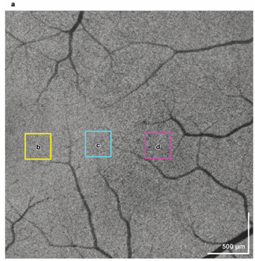
According to Professor Oh, this system can show microstructures of multiple layers of retinal nerve fibers and photoreceptors at cellular resolution in just 2.3 seconds. This high-resolution retinal imaging technology has significant implications for expanding pathophysiological knowledge of ocular diseases and their treatment. The team's findings may pave the way for the development of more accessible and affordable treatment with its novel postprocessing algorithm and cost-efficient diagnosis device for diagnosing and treating eye diseases.